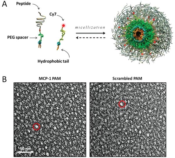
Figure 1.
Design and structure of MCP-1 PAMs. A) Schematic depicting PAM self-assembly. PAs consist of a distearoyl hydrophobic tail (two 18-carbon chains) and a PEG spacer that was conjugated to the MCP-1 peptide that corresponds to the CCR2-binding motif (residues 13–35), scrambled peptide, or the Cy7 fluorophore. Fluorescently-labeled, mixed micelles consisted of peptide-containing and Cy7-labeled amphiphiles in a 90:10 molar ratio. B) Representative TEM images of MCP-1 PAMs (L) and scrambled PAMs (R). Both micelles are spherical in shape with a diameter on the order of 10 nm.

