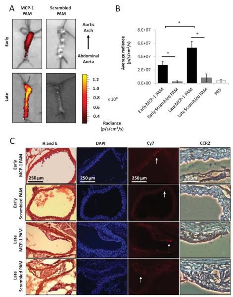
Figure 3.
MCP-1 PAMs detect and discriminate between stages of atherosclerosis in vivo. Optical imaging of early- and late-stage atherosclerotic aortas of ApoE knock-out mice injected with A) MCP-1 PAMs or scrambled PAMs after 24 h. Representative aortas are shown. B) Quantification of micelle binding. Data points are mean ± SD, *P < 0.05, N ≥ 3 mice. C) Representative histological and immunohistochemical staining of cross-sections derived from early- and late-stage atherosclerotic aortas treated with MCP-1 or scrambled PAMs. From left to right: H and E, DAPI, Cy7 (PAM) signal, and magnified regions showing CCR2 expression via DAB staining (brown). Arrows in the Cy7 images denote the region that was magnified in corresponding CCR2 image.

