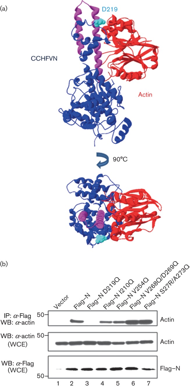
Fig. 8.

Molecular modelling of the CCHFV N–actin interaction. (a) The model was constructed with the structures of CCHFV N protein PDB: 4AQF (Wang et al., 2012) (in blue) and human actin PDB: 1ATN (in red), using the program PatchDock as described in Methods. The coiled-coil motif is in pink. The aspartate (D) in position 219 is highlighted in cyan in the CCHFV N protein structure. (b) HEK 293T cells were transfected with CCHFV N protein or N protein Flag-tagged point mutants, as indicated. After 24 h, the cells were harvested and lysed, and the overexpressed proteins were immunoprecipitated with anti-Flag (IP: α-Flag). After SDS-PAGE, expression levels of WT Flag–N, each N protein mutant and actin were examined by Western blotting (WB) using the antibodies indicated. Molecular masses of markers are indicated on the left in kDa. WCE, Whole-cell extracts.
