
Figure 5.
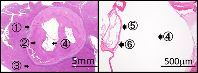
Histopathology showing an alveolar echinococcosis cyst of 3.7×4.1 mm in size. ①: Leucocyte layer, ②: fibrous layer, ③: normal liver tissue, ④: multilocular cysts, ⑤: laminated layer and ⑥: germinal layer.
Official websites use .gov
A
.gov website belongs to an official
government organization in the United States.
Secure .gov websites use HTTPS
A lock (
) or https:// means you've safely
connected to the .gov website. Share sensitive
information only on official, secure websites.
Histopathology showing an alveolar echinococcosis cyst of 3.7×4.1 mm in size. ①: Leucocyte layer, ②: fibrous layer, ③: normal liver tissue, ④: multilocular cysts, ⑤: laminated layer and ⑥: germinal layer.