
Figure 1.
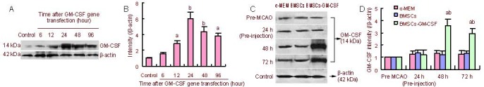
GM-CSF expression in GM-CSF-transfected BMSCs and the effect of BMSCs-GM-CSF on GM-CSF production in vivo.
(A) GM-CSF protein content in BMSCs was examined by western blot assay at 6, 12, 24, 48, and 96 hours after GM-CSF gene transfection.
(B) Bar graph shows GM-CSF protein levels at different time points after GM-CSF gene transfection in BMSCs. Data are expressed as mean ± SD. aP < 0.05, bP < 0.01, vs. control group, two-way analysis of variance followed by post-hoc Tukey test. The preMCAO group was standardized to 1.
(C) GM-CSF protein content was examined by western blot assay using protein samples from the ischemic hemisphere of rats treated with α-MEM, BMSCs, and BMSCs-GM-CSF at 24, 48, and 72 hours after MCAO and before MCAO. At 24 hours after MCAO, the right lateral corpus striatum was injected with BMSCs or BMSCs-GM-CSF. In the image, 24 h after MCAO refers to results before injection. 48, 72 hours after MCAO refer to 24, 48 hours after injection.
(D) Bar graph shows GM-CSF protein levels in the ischemic hemisphere of rats treated with α-MEM, BMSCs, and BMSCs-GM-CSF at 24, 48, and 72 hours after MCAO and before MCAO. Data are expressed as mean ± SD. aP < 0.01, vs. control (α-MEM) group; bP < 0.01, vs. BMSCs group, two-way analysis of variance followed by post-hoc Tukey test. The pre-MCAO group was standardized to 1.
GM-CSF: Granulocyte-macrophage colony-stimulating factor; BMSCs: bone marrow stromal cells; MCAO: middle cerebral artery occlusion.