
Figure 1.
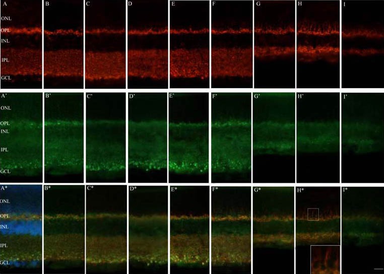
Fluorescence immunoreactivity of synaptophysin (SYN) and synapse-associated protein 102 (SAP102) following acute high intraocular pressure (HIOP) in the rat retina under a fluorescence microscope.
Panels A–I depict SYN (red) antibody labeling in the normal control, sham surgery, and HIOP 2 hours, 6 hours, 12 hours, 1 day, 3 days, 7 days and 14 days groups, respectively. Panels A’–I’ indicate SAP102 (green) antibody labeling in the normal control, sham surgery, and HIOP 2 hours, 6 hours, 12 hours, 1 day, 3 days, 7 days and 14 days groups, respectively. Panels A*–I* show double-labeling of SYN (red) and SAP102 (green) antibody in the normal control, sham surgery, and HIOP 2 hours, 6 hours, 12 hours, 1 day, 3 days, 7 days and 14 days groups, respectively. Nuclei of the cells were marked with Hoechst in A* (blue). The rectangle below in H* is a higher magnification of the upper selected region.
ONL: Outer nuclear layer; OPL: outer plexiform layer; INL: inner nuclear layer; IPL: inner plexiform layer; GCL: ganglion cell layer. Scale bar represents 25 μm.