
Figure 2.
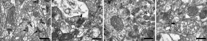
Radiation-induced ultrastructural disarray in the hippocampal CA1 neuropil of Wistar rats (transmission electron microscopy, scale bars: 0.5 μm).
(A) The stratum radiatum of the hippocampal CA1 region from a sham-irradiated rat shows an abundance of tightly packed neuro-glial profiles with numerous synaptic contacts (arrows).
(B) The stratum radiatum of the hippocampal CA1 region from an irradiated rat from a different litter shows a striking scarcity of synaptic contacts (synaptic contacts are indicated by arrow) and a gross loss of neuro-glial profiles.
(C) Mitochondria in the hippocampal CA1 region of sham-irradiated rats appear healthy with clear double membranes and tight, orderly cristae.
(D) Mitochondria in the hippocampal CA1 region of irradiated rats are swollen with balloon-like cristae (arrow).