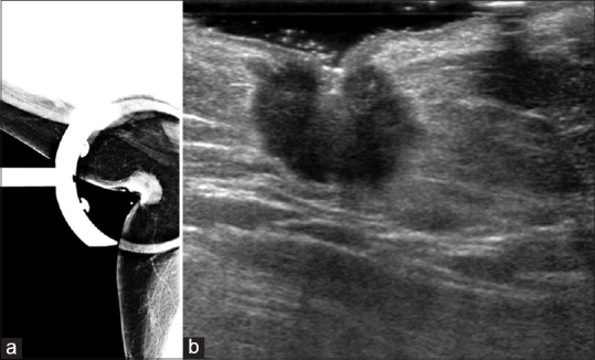
Figure 1.

(a) Spot compression mammographic image of the right axilla of our patient shows a round hyperdense mass. (b) Ultrasound image of the right axilla shows an irregular, hypoechoic mass. On biopsy, this was found to be an invasive ductal carcinoma
