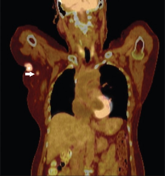
Figure 2.

Fused coronal PET-CT image of our patient before the axillary lymph node biopsy shows a lobulated FDG-avid mass (star) in the right axilla, which represents the patient's known axillary invasive ductal carcinoma. An adjacent small FDG-avid round structure (arrow) was suspicious for metastatic axillary lymphadenopathy
