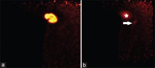
Figure 3.

(a) PEM imaging of the right axillary tail shows the FDG-avid mass in the right axilla, which represents the patient's known invasive ductal carcinoma (IDC). Even with an approximate 37 MBq activity of 18F-FDG in the patient's system, the patient's carcinoma is well-visualized. (b) Another axillary tail postbiopsy PEM image of the right axilla shows the carcinoma (star) and the FDG-avid axillary lymph node (arrow). In comparison to the whole-body positron emission tomography-computed tomography image [Figure 2], the lymph node has changed in size and configuration, indirectly indicating correct biopsy sampling
