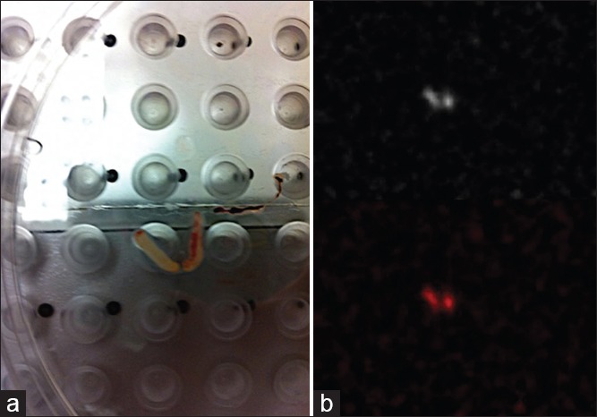
Figure 4.

(a) Photograph of the ultrasound core needle biopsy specimen positioned on the detector plate of the PEM scanner. (b) Corresponding PEM image of the of the specimen reveals FDG activity within the core samples, confirming successful sampling of the lymph node
