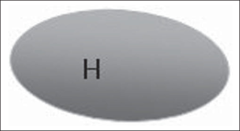
Figure 1.
Bedi type 1 lymph node. Without visible cortex. (H, hilum).
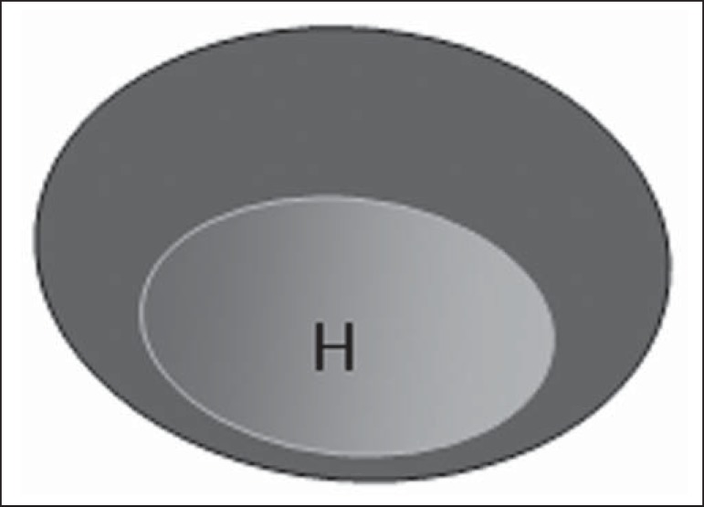
Figure 2.
Bedi type 2 lymph node. Uniform cortex ≤ 3 mm. (H, hilum).
Figure 3.
Bedi type 3 lymph node. Uniform cortex > 3 mm cortex. (H, hilum).
Figure 4.
Bedi type 4 lymph node. Entirely lobulated cortex. (H, Hilum).
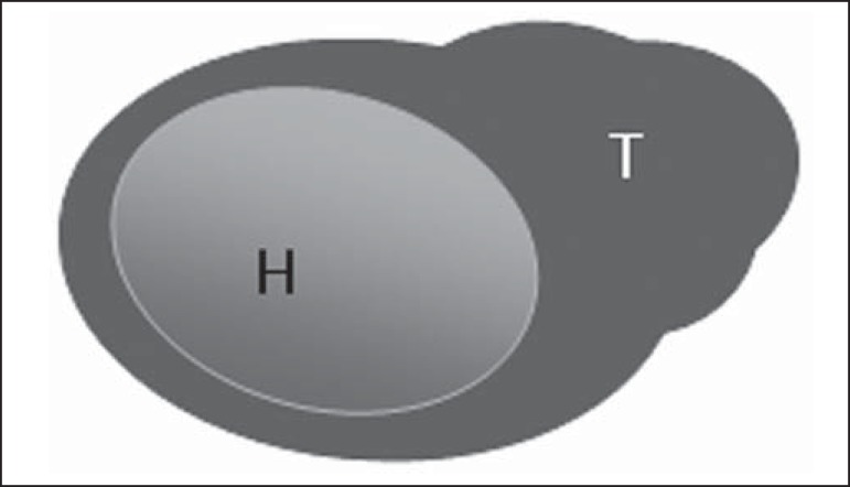
Figure 5.
Bedi type 5 lymph node. Cortex with focal lobulation. (H, hilum; T, tumor cell deposit).
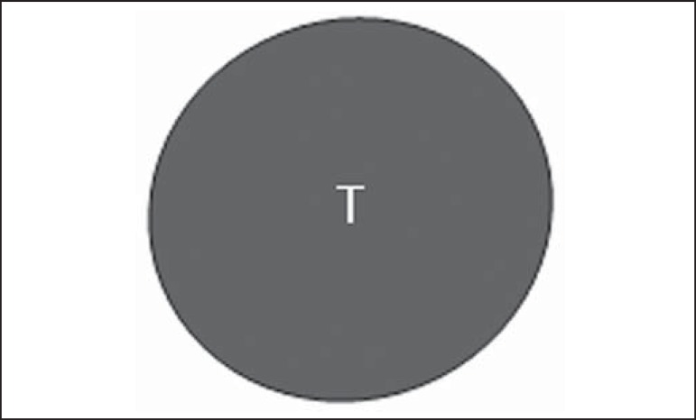
Figure 6.
Bedi type 6 lymph node. Completely hypoechogenic lymph node, absent hilum (T, tumor cell deposit).
Figure 7.
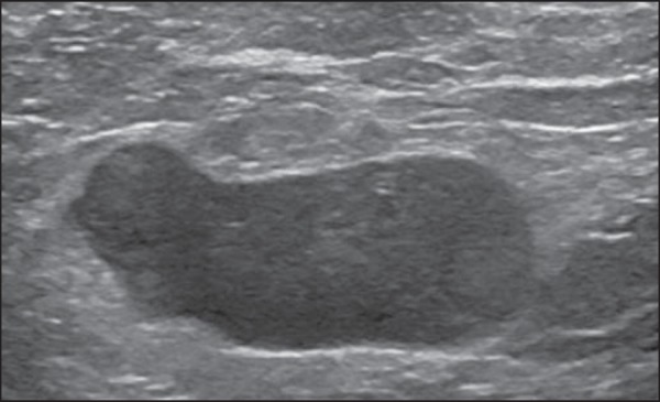
Bedi type 6 lymph node at ultrasonography. Lymph node without an apparent hilum.

