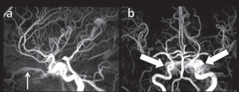
Figure 7.
MR angiography of intracranial vessels. a: Lateral view. b: Anteroposterior view. Female 24-year-old patient with diagnosis of spontaneous CCF. Contrast enhancement of the left cavernous sinus which is dilated, with reflux into the right CS (bold arrows). Early contrast enhancement of the SOV (thin arrow) which is dilated.

