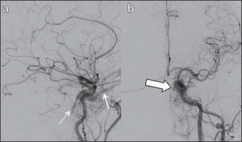
Figure 8.
Digital subtraction cerebral angiography, arterial phase. a: Lateral view. b: Anteroposterior view. Female 69-year-old patient with a diagnosis of DCSF. Early CS contrast enhancement of the cavernous sinus (bold arrow). Observe the dilatation and early enhancement of the SOV (thin arrow). This CS fistula presented anterior drainage into the SOV and posterior drainage into IPS (dashed arrow). Observe the presence of multiple small supplying meningeal branches (bold arrow)

