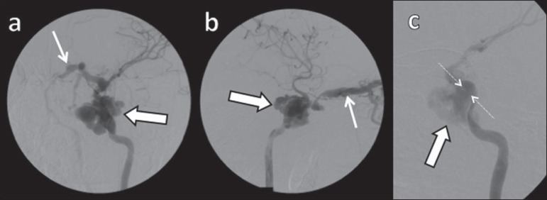
Figure 10.
Digital subtraction cerebral angiography, arterial phase. a: Left anterior oblique view. b: Lateral view. c: Working view. Male 24-year-old patient with a diagnosis of post-traumatic CCF. Early contrast enhancement of the CS (bold arrows), which is dilated. Early drainage into ectatic SOV (thin arrows). Observe the exact point of laceration of the ICA which communicates with the CS (between the dotted arrows).

