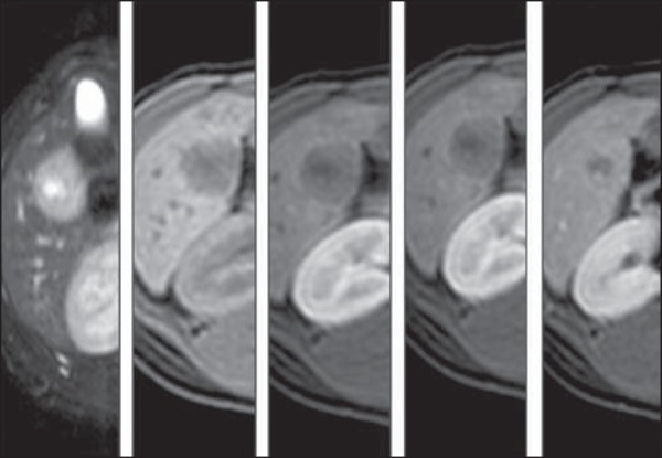
Figure 1.

Magnetic resonance imaging (T2 STIR, TIGRE, pre-contrast, arterial, portal and equilibrium phases). Circumscribed nodule located in the periphery of the visceral aspect of the segment IV, with target sign on T2-weighted sequence, low signal intensity on T1-weighet sequence and hypovascular and progressive contrast enhancement. The lesion margins are indistinguishable at the delayed phase.
