Figure 3.

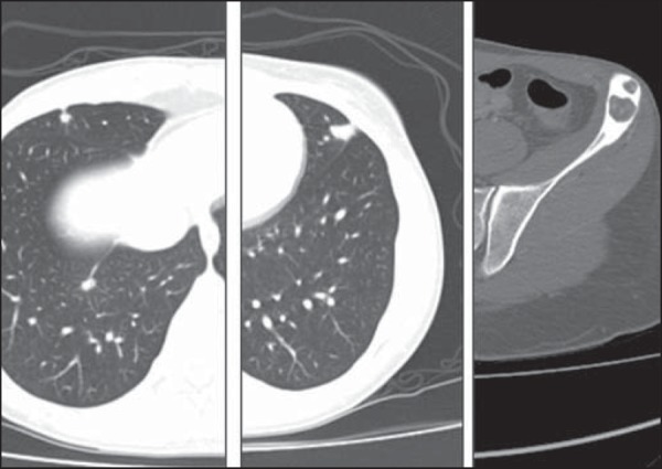
Computed tomography. Pulmonary nodules located at the periphery of the costal surface of the middle lobe and in the lingular segment. Lytic bone lesions with marginal sclerosis in the left iliac bone.

Computed tomography. Pulmonary nodules located at the periphery of the costal surface of the middle lobe and in the lingular segment. Lytic bone lesions with marginal sclerosis in the left iliac bone.