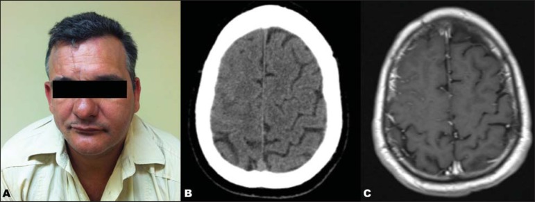
Figure 1.
A: Right hemifacial alteration characterized by remarkable atrophy and deformity. B: Non-contrast-enhanced, axial, cranial CT demonstrating fading of the sulci in the right frontal lobe. C: Axial MRI, contrast-enhanced T1-weighted sequence more clearly demonstrating the fading of the sulci as well as the associated meningeal enhancement.

