
Figure 2:
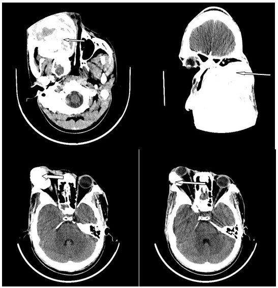
Enhanced axial and coronal images of the craniofacial bone in a 31-year-old male showing an expansile and destructive mass of the right maxillary bone (arrows) with compression of the orbit, nasal cavity and oral cavity. The histology diagnosis was fibromyxoma, which is a benign tumour.