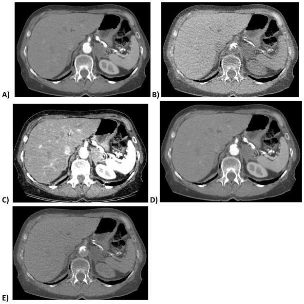
Figure 1. 70 year old woman with lipid-rich left adrenal adenoma.
DECT images show the nodule in the arterial phase of enhancement (A) and a virtual non-contrast (VNC) image (B) derived from the same series using the iodine:water material basis pair. Calcium in the aorta remains in image B after iodine subtraction. Images C–E are virtual monochromatic spectral (VMS) images at 40, 75 and 140 keV, respectively. Note the decreasing conspicuity of iodine at higher reconstructed monochromatic energy levels. E is similar but not identical to B, with E exhibiting persistent subtle corticomedullary differentiation and less noise.

