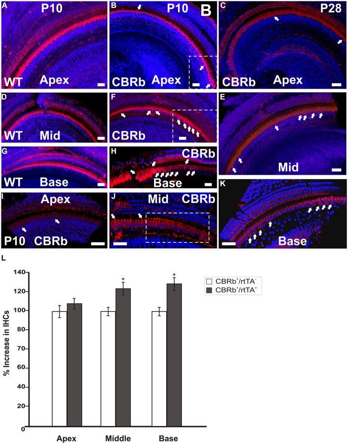
FIGURE 11.
Immunohistochemical detection of myosin VIIa (M7a)-positive supernumerary cells in the cochleae of DN-CBRb+/ROSA-CAG-rtTA+ (CBRb) at postnatal (P) day 10 and P28. Compared to the DN-CBRb+/ROSA-CAG-rtTA- (WT) control, which show a single line of cells at the IHCs’ region throughout the length of the cochlea (A,D,G), P10 (B,E,H–J) and P28 (C,F,K) CBRb mutants exhibited extra M7a-positive cells (arrows and boxed regions), which were more concentrated in the basal and middle turns of the cochlea. Of note, the density of supernumerary cells was higher in P10 ears compared to P28. A close up of boxed areas in (B,E) are shown in (I) and (J), respectively. Boxed area in (J) highlights an area where IHCs are seen in as a double row. The blue background in all figures corresponds to DAPI staining. Bar = 10 μm. (L) Counting of Myosin VIIa-positive cells at the IHCs’ region of CBRb mice cochleae at P10 revealed a significant increase in cells, particularly at the middle and basal turns, when compared to DN-CBRb-/ROSA-CAG-rtTA+ mice. Error bars show SEM. *P < 0.05.

