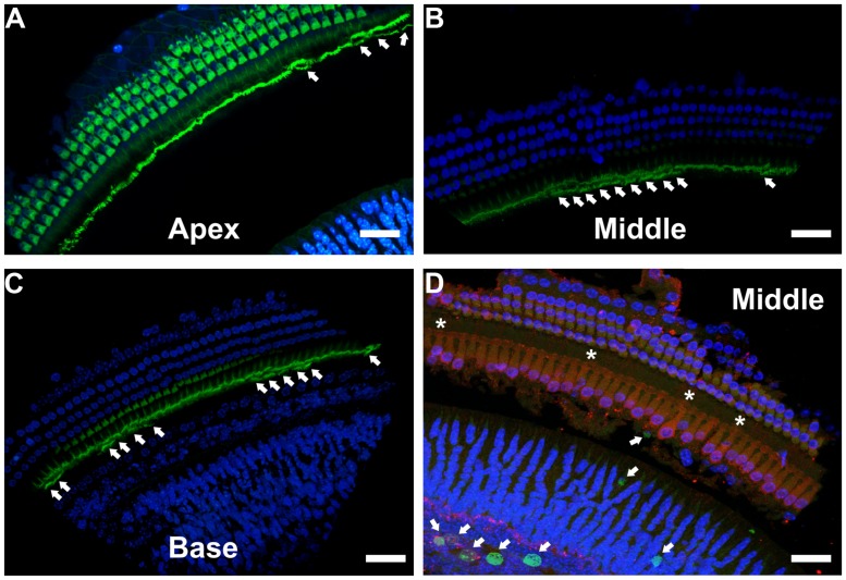
FIGURE 12.
Immunohistochemical localization of stereocilia and proliferating cells on DN-CBRb+/ROSA-CAG-rtTA+ mouse cochleae. (A–C) Phalloidin (green) staining of P10 CBRb+/ROSA-CAG-rtTA+ mouse cochlea revealed the presence of F-actin-positive stereocilia-like structures (arrows). Due to the peculiar cochleae anatomy, (B,C) were rotated to facilitate visualization of the stereocilia. (D) EdU (green) labeling in DN-CBRb+/ROSA-CAG-rtTA+ (CBRb) at P10 showed no proliferation within the OC; however, a few EdU-positive cells were observed at the greater epithelial ridge. Asterisks indicate supernumerary cells. DAPI = Blue; Myosin VIIa = Red. Bar = 10 μm.

