
Fig. 8.
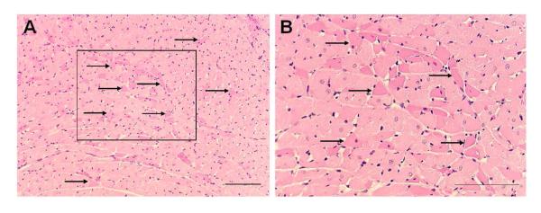
Eosinophilia of discrete cardiac myocytes (indicated by arrows) was seen diffusely at low (A) and high (B) power magnification in sections of left ventricular myocardium stained with hematoxylin and eosin. The box in A is seen at higher magnification in B. The scale bars = 50 μm. From Nayate et al as permitted (Nayate et al., 2008).