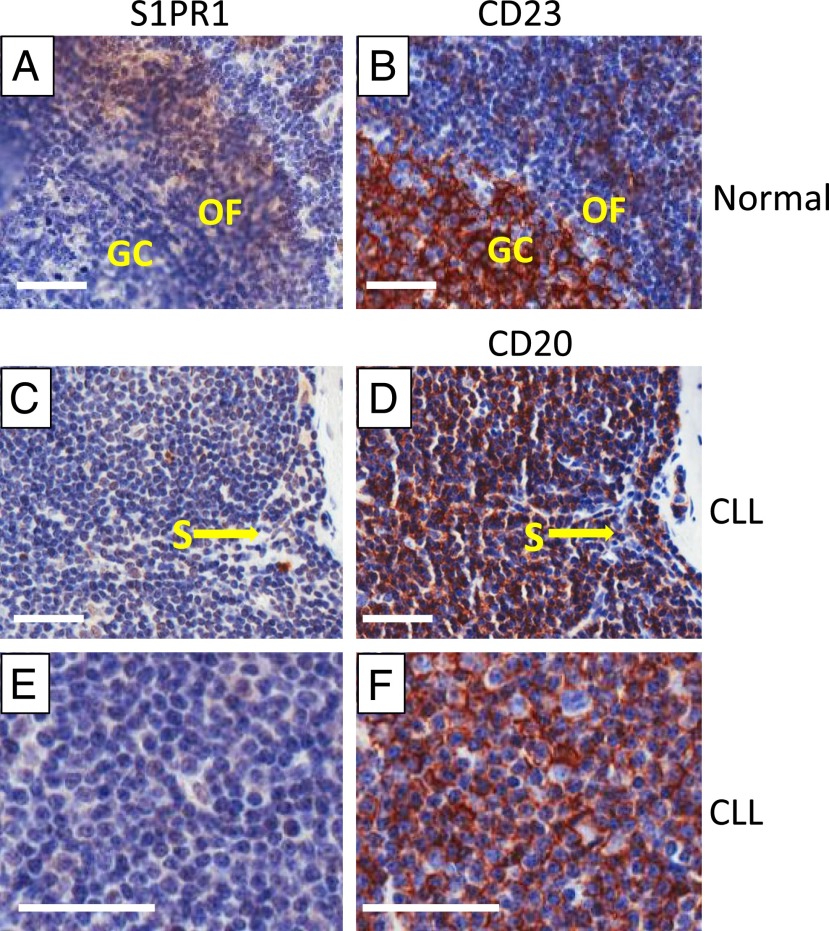
FIGURE 1.
Expression of S1PR1 in normal and CLL node. (A and B) Normal lymph node. The cells of the germinal center (GC) are identified by staining for CD23, which is mainly expressed on activated B cells. Staining of the adjacent section for S1PR1 shows that CD23+ cells do not express S1PR1, whereas the cells within the outer follicle (OF) stain positively. (C–F) Sequential section of CLL lymph nodes stained for S1PR1 and CD20. In (C) and (D), it can be clearly seen that the endothelial cells lining the sinus (S) and other endothelial cells express S1PR1, whereas CD20+ CLL cells lack the receptor. (E) and (F) show a representative proliferation center in a CLL lymph node. CD20+ CLL cells do not express S1PR1. Parallel staining with the relevant isotypic control Ab is shown in Supplemental Fig. 2A. Scale bar, 50 μM; original magnification ×20.

