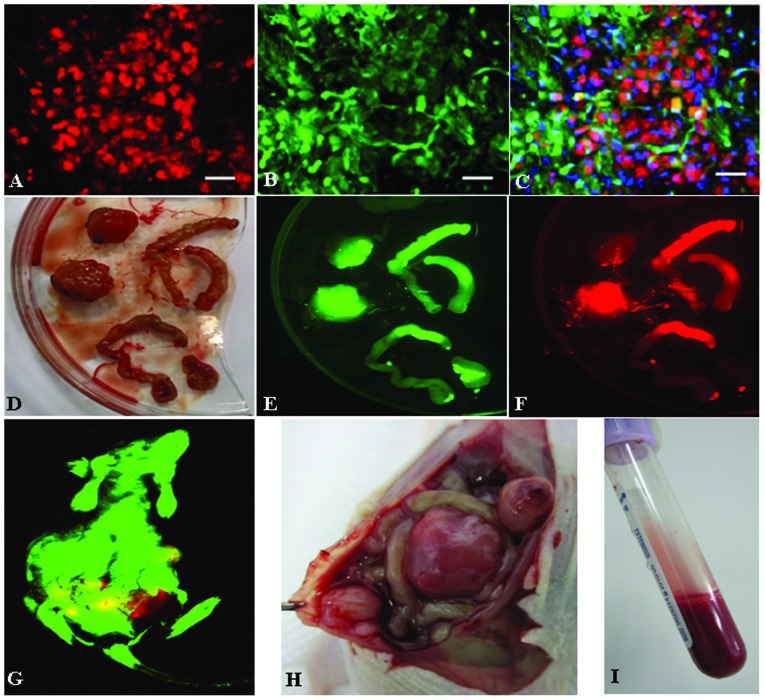
Figure 2.
Double-color fluorescent tracer effects of SU3-red fluorescent protein (RFP)/green fluorescent protein (GFP) tumor model. (A–C) Cryosections of abdominal transplanted tumor under confocal microscopy (scale bar=20 μm); (D) Abdominal tumor nodules and the small intestine invaded by tumor cells; (E–F) D in living imaging system; (G) Tumor-bearing mice in living imaging system; (H) Tumor nodules in abdominal anatomy of tumor-bearing mice; (I) Intraperitoneal bloody ascites from tumor-bearing mice.

