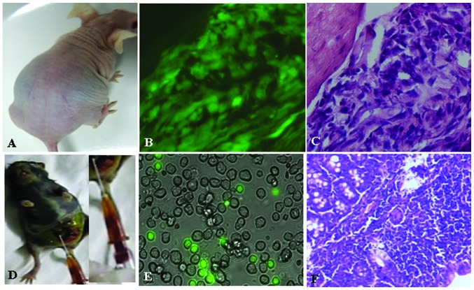
Figure 6.
Tumorigenicity tests in SU3-induced host celiac tumor cells (SU3-ihCTCs). (A) SU3-ihCTCs were transplanted into the abdomen of BALB/c nude mice, leading to malignant ascites and abdominal distension; (B) Fluorescence microscope image of cryosections of SU3-ihCTC abdominal implantation (magnification, ×400); (C) Hematoxylin and eosin (H&E) staining of tissue adjacent to B (magnification, ×400); tumor cells exhibited significant pleomorphism and were densely arranged; (D) RAW264.7 was inoculated into green fluorescent protein (GFP) nude mice intraperitoneally, forming bloody ascites and tumor nodules; (E) Fluorescence microscope image (magnification, ×400), RAW264.7-derived cells and host-derived GFP-expressing green-colored cells were observed in the ascites; (F) Optical microscope image of sections of GFP nude mice with RAW264.7 intraperitoneal inoculation (H&E staining, ×100).

