
FIG 7.
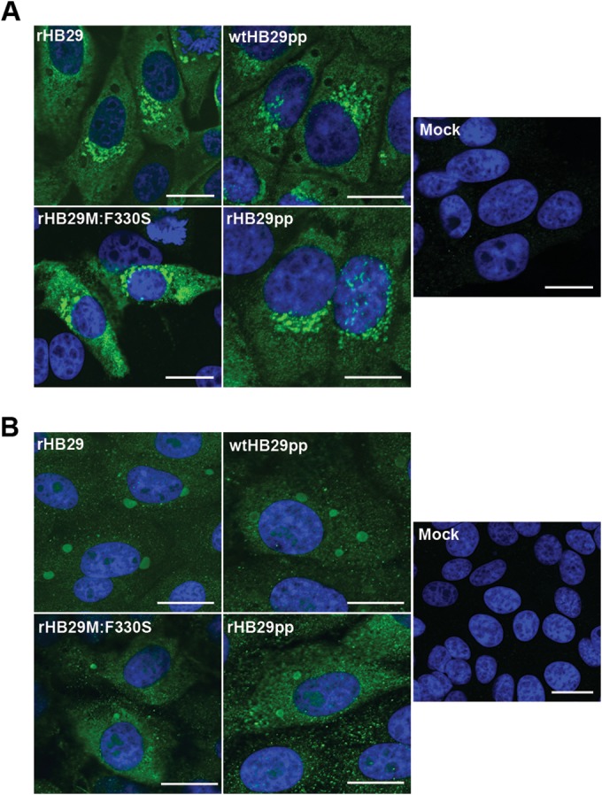
Intracellular localization of N and NSs proteins in infected cells. Vero E6 cells were infected at an MOI of 5 with rHB29, wtHB29pp, rHB29pp, or rHB29M:F330S or mock infected. At 24 h p.i., the cells were fixed with 4% formaldehyde, followed by staining with either anti-N (A) or anti-NSs (B) monospecific antibodies. Samples were counterstained with DAPI. Cells were examined with a Zeiss LSM 710 confocal microscope. Bar, 10 μm.