
FIG 1.
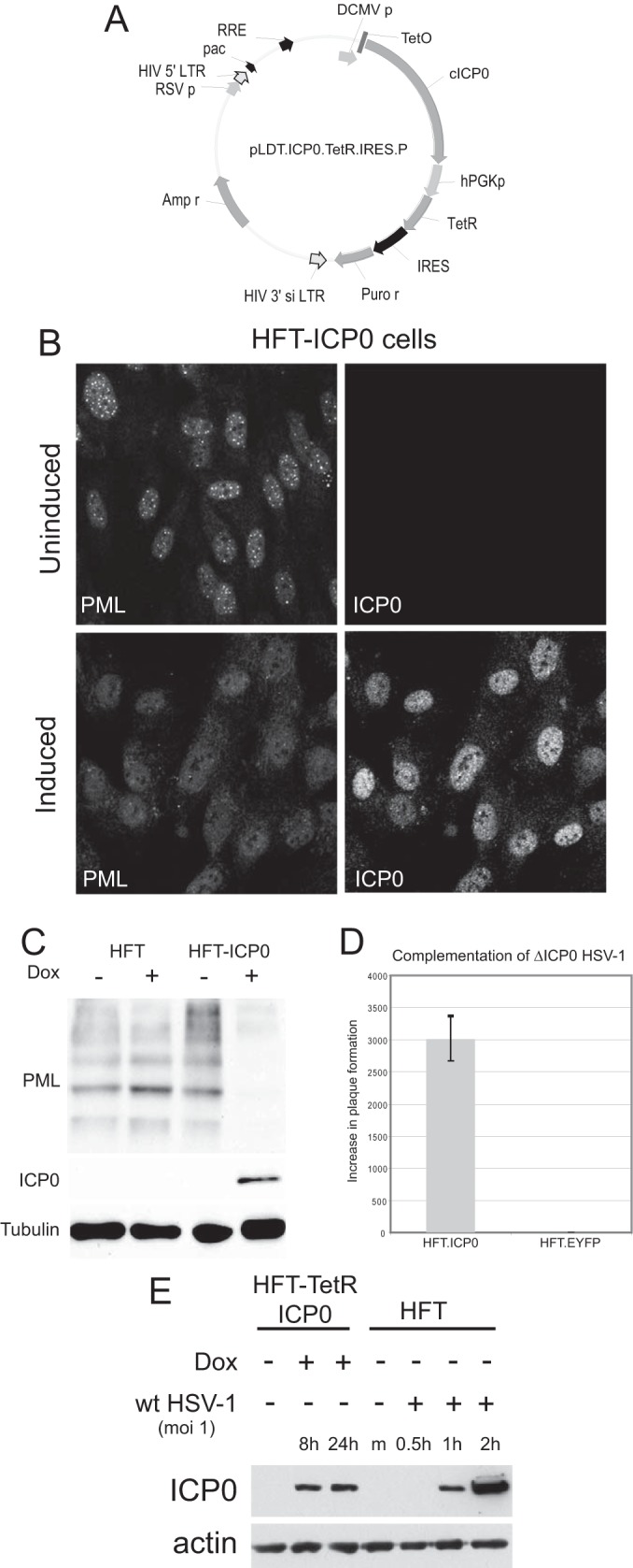
Characterization of a system for inducible expression of ICP0 in human fibroblasts. (A) A map of lentivirus plasmid vector pLDT.ICP0.TetR.IRES.P. The key features of the lentivirus vector are noted: pac, HIV packaging sequence; RSV p, RSV promoter; RRE, Rev response element; hPGK, human phosphoglycerate kinase promoter; IRES, internal ribosome binding site; Puro r, puromycin resistance coding sequence; LTR, long terminal repeat. (B) Immunofluorescence staining of ICP0 and PML in HFT-ICP0 cells before and after induction of ICP0 expression. (C) Western blot analysis of HFT.ICP0 cells, probing for PML, ICP0, and tubulin. (D) Complementation of ICP0 null mutant HSV-1 in plaque formation assays. HFT.ICP0 and HFT.EYFP cells were treated with doxycycline (100 ng/ml) for 24 h before infection with dl1403/CMV lacZ, an ICP0 deletion virus carrying a β-galactosidase marker gene. Plaques were counted 24 h after infection by staining for β-galactosidase. The results are expressed as fold increase in PFU per ml in each cell type compared to HFT cells; error bars are ± standard deviations. EYFP, enhanced yellow fluorescent protein. (E) Comparison of the level of ICP0 expressed after 8 and 24 h of induction of HFT-TetR-ICP0 cells (made by sequential transduction with TetR and ICP0 lentiviral vectors) with that expressed during the early stages of wt HSV-1 infection of HFT cells (MOI of 1; based on the titer of wt HSV-1 on HFs). Dox, doxycycline; m, mock infected.