FIG 3.
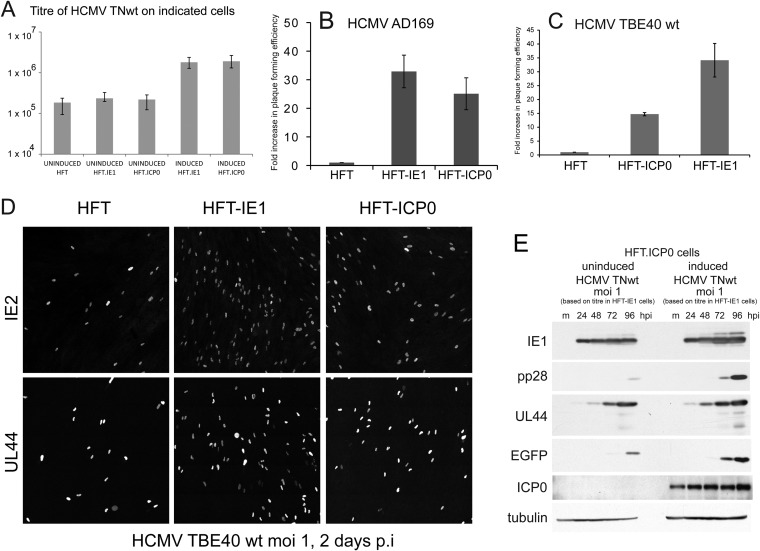
ICP0 and IE1 augment infection of wt HCMV. (A to C). Stocks of wt HCMV strains TNwt (A), AD169 (B), and TBE40 wt (C) were titrated on HFT, HFT-ICP0, and HFT-IE1 cells. In panel A the results are presented as the mean of the average of three independent experiments, with the error bars indicating the range of values obtained, plotted on a log10 scale. In panels B and C, the results are presented as fold increases above the titers in HFT cells (mean ± standard deviation). (D) Comparison of the proportion of IE2- and UL44-positive cells in HFT, HFT-IE1, and HFT-ICP0 cells with TBE40 as indicated. p.i., postinfection. (E) Western blot analysis of HCMV TNwt infection on HFT.ICP0 cells with or without induction of ICP0 expression 24 h before infection. Samples were collected at 24, 48, 72, and 96 h after infection then probed with anti-IE1 MAb E13, anti-pp28 MAb, anti-UL44 MAb, anti-EGFP rAb, anti-ICP0 MAb 11060, and anti-tubulin MAb, as indicated. hpi, hours postinfection.

