FIG 4.

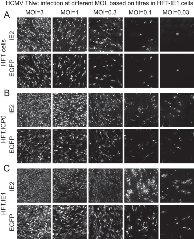
Analysis of HCMV TNwt infection in HFT, HFT-ICP0, and HFT-IE1 cells by immunofluorescence. Cells were infected at decreasing multiplicities, as indicated, and then examined for IE2 and EGFP expression at 5 days after infection.

Analysis of HCMV TNwt infection in HFT, HFT-ICP0, and HFT-IE1 cells by immunofluorescence. Cells were infected at decreasing multiplicities, as indicated, and then examined for IE2 and EGFP expression at 5 days after infection.