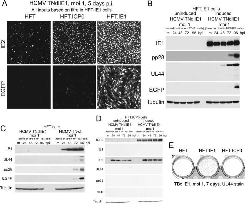
FIG 5.
IE1 but not ICP0 complements IE1 mutant HCMV infection. (A) Analysis of IE2 and EGFP expression in HFT, HFT-ICP0, and HFT-IE1 cells infected with HCMV TNdlIE1. p.i., postinfection. (B) Western blot analysis of pp28, UL44, and EGFP expression at the indicted time points after HCMV TNdlIE1 infection of uninduced and induced HFT.IE1 cells. Expression of these proteins from the viral genome was not detectable in induced HFT-ICP0 cells (data not shown). Tubulin provides the loading control. (C) Comparison of viral gene expression by TNdlIE1 and TNwt after infection of HFT cells. (D) Analysis of viral gene expression after infection of uninduced and induced HFT-ICP0 cells with TNdlIE1. (E) Comparison of UL44 expression by TBdlIE1, the IE1 mutant derived from HCMV strain TBE40, in induced HFT, HFT-IE1, and HFT-ICP0 cells as detected by immunohistochemistry staining of infected cell cultures.

