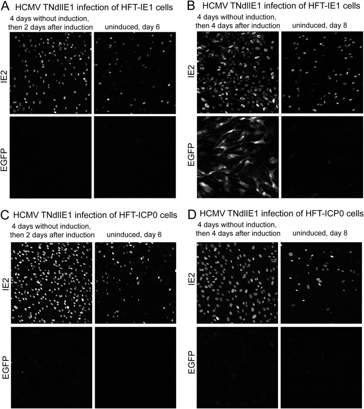
FIG 9.
Stimulation of HCMV gene expression after delayed expression of IE1 during HCMV TNdlIE1 infection of HFT-IE1 cells. Cells were infected with HCMV TNdlIE1 (MOI of 1, based on the titer in HFT-IE1 cells) prior to induction and then incubated for a further 4 days. At that point, doxycycline was added to 100 ng/ml to induce expression of IE1; then samples were analyzed for expression of IE2 and EGFP at various times thereafter. The results shown in panels A and B illustrate the basal level of IE2 expression in the absence of IE1, the increase in the number of cells expressing IE2 after induction, and the progression of these cells to EGFP expression in the presence of induced IE1 (B). Panels C and D show images from an analogous experiment using HFT-ICP0 cells. The images are composites of 9 tiled images captured with a 40× lens at a zoom factor of 1.

