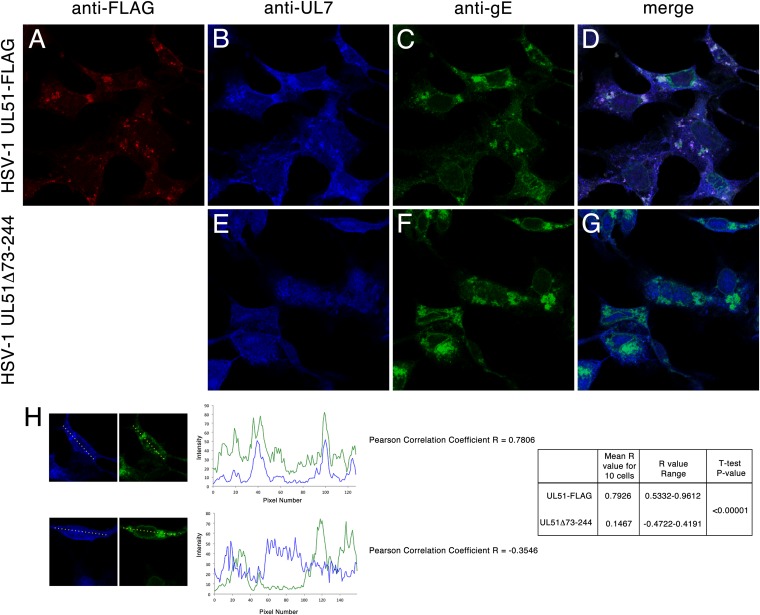
FIG 3.
Localization of pUL51, pUL7, and gE in infected cells. Vero cells infected for 14 h with HSV-1 UL51WT-FLAG (A to D) or HSV-1 UL51Δ73-244 (E to G) were fixed and probed with anti-FLAG antibody to detect pUL51 (A and D), anti-UL7 antiserum (B, D, E, and G), and mouse monoclonal antibody directed against gE (C, D, F, and G). Confocal images of single z-sections taken near the center of the nucleus (i.e., where the nuclear cross-section is the largest) with a 60× oil objective are shown. Negative controls (not shown) were normal rabbit serum for anti-UL7 rabbit antiserum and uninfected cells for anti-gE and anti-FLAG and showed no fluorescence at the laser and detector settings used to obtain these images. (H) Quantitation of gE and pUL7 colocalization. gE and pUL7 fluorescent intensities were measured across the linear profiles of 10 randomly selected cells, as described in Materials and Methods. Representative profiles from one cell infected with UL51-FLAG (top) and UL51Δ73-244 (bottom) are shown along with the profile plots and derived Pearson correlation coefficients. Aggregate results from 10 profiles are shown at the right.

