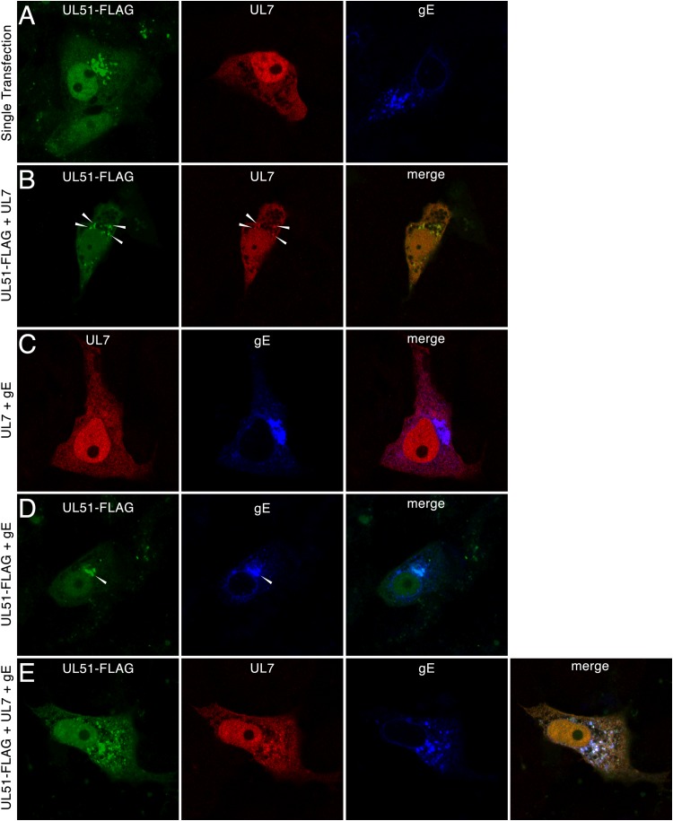
FIG 4.
Localization of pUL51, pUL7, and gE in transfected cells. Vero cells were transfected with plasmids for 24 h and then fixed and probed by indirect immunofluorescence. Images of single z-sections taken near the center of the nucleus with a 60× oil objective are shown. Primary antibodies are indicated at the tops of the panels, and the plasmids used for transfection are indicated to the left of each row. White arrowheads, areas of colocalization between pUL51 and pUL7.

