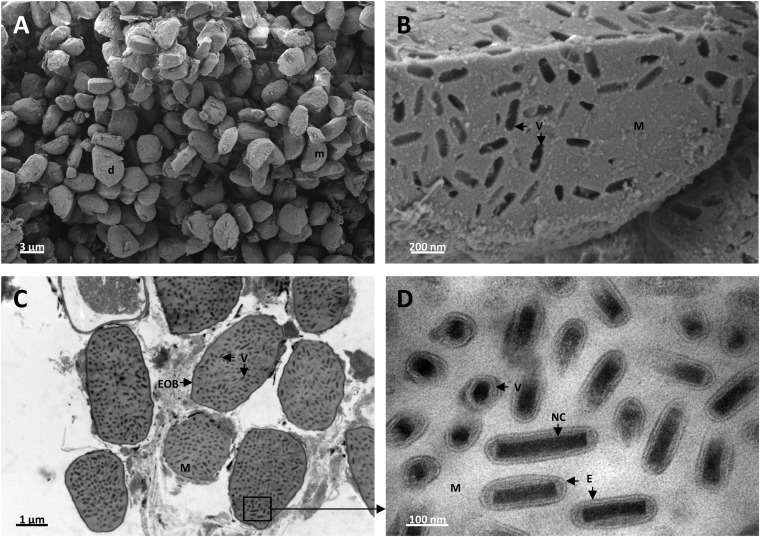
FIG 8.
Occlusion bodies, virions, and nucleocapsids from T. oleracea purified nudivirus visualized by scanning (A and B) and transmission (C and D) electron microscopies. (A) Purified ToNV OBs shaped irregularly from droplet (d) to moon (m) by way of all conceivable ovoid and ellipsoid forms. (B) Section of typical OB enlarged image showing rod-shaped virion (V) prints inside the protein matrix (M). (C) Thin cross-section of enveloped occlusion bodies (EOB) filled with numerous virions (V) and protein matrix (M). (D) Enlarged image of protein matrix (M)-embedded virions (V) displaying, mainly in cross-sectional and longitudinal views, nucleocapsids (NC) surrounded by single bilayer membranes forming the envelopes (E).

