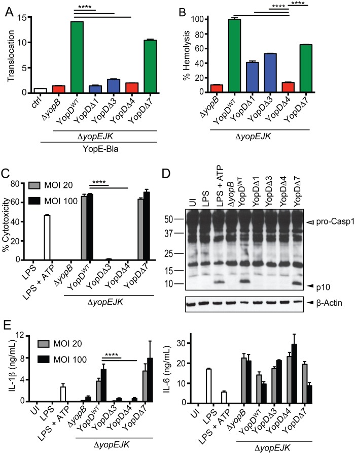
FIG 1 .
Translocation is required for inflammasome activation by Yersinia T3SS. (A) HeLa cells were infected with indicated strains expressing a YopE–β-lactamase fusion protein (YopE-Bla) or ΔyopEJK mutant lacking the β-lactamase construct (ctrl). Cells were loaded with CCF4-AM dye, and the ratio of blue to green signal (translocation) was calculated as described in Materials and Methods. Results are representative of 3 independent experiments. (B) Sheep red blood cells were infected with indicated YopD deletion mutants or the ΔyopB and ΔyopEJK controls. Supernatants were assayed for release of hemoglobin as described in Materials and Methods. The graph is representative of two to four independent experiments with 6 independent replicates per sample. (C) BMDMs were infected with indicated YopD deletion mutants (ΔyopB or ΔyopEJK) or treated with LPS or LPS plus ATP, and cytotoxicity was determined by LDH release. The graph is of a representative experiment from one of five independent experiments (MOI, 20) or 2 independent experiments (MOI, 100) with 3 replicates per sample. (D) BMDMs were infected with the indicated bacterial strains or treated with LPS or LPS plus ATP. Cell lysates were assayed for p10 antibody label, processed caspase-1 (pro-Casp1), and actin as indicated. (E) Supernatants from BMDMs infected with indicated bacterial strains were assayed for levels of secreted IL-1β and IL-6 by ELISA as described in Materials and Methods. UI, uninfected. ****, P < 0.0001.

