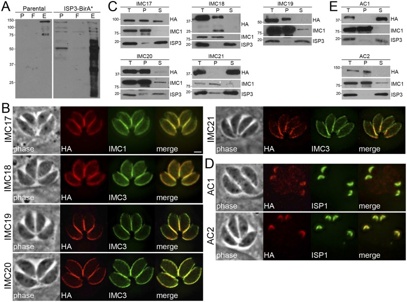
FIG 2 .
Identification of novel IMC proteins by ISP3 BioID. (A) Western blot showing enrichment of biotinylated proteins from parental and ISP3-BirA* lysates by streptavidin magnetic beads, as assessed by monitoring of the precolumn (P), flowthrough (F), and elution (E) fractions. (B) ISP3 BioID hits were localized to the IMC by endogenous tagging, where they colocalize with the IMC markers IMCs 1/3. Similar to ISP3-BirA*, these proteins stain the central and basal subcompartments of the IMC but not the apical cap. Red, mouse or rabbit anti-HA antibodies; green, mouse anti-IMC1 or rat anti-IMC3 antibodies. Scale bar = 2 µm. (C) Western blots showing detergent extraction analysis of ISP3 BioID hits. The total lysate (T) was partitioned into the insoluble pellet (P) or soluble (S) fractions. Fractionation was monitored using IMC1 (insoluble) and ISP3 (soluble) controls. (D) IFA showing two additional ISP3 BioID hits that localize to the apical cap demarked by ISP1. Red, rabbit anti-HA antibody; green, mouse anti-ISP1 antibody. (E) Detergent extractions of ACs 1/2 as described in part C.

