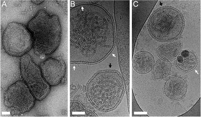
FIG 1 .
HPIV3 imaged by negative-stain (A) and cryo- (B and C) electron microscopy. As is particularly evident in cryo-electron micrographs, nearly all particles were rounded rather than filamentous in morphology. In addition to a complex, dense layer of surface density (black arrows), regions of ordered density (white arrows) were evident, both attributed to the viral glycoproteins. Scale bars, 100 nm.

