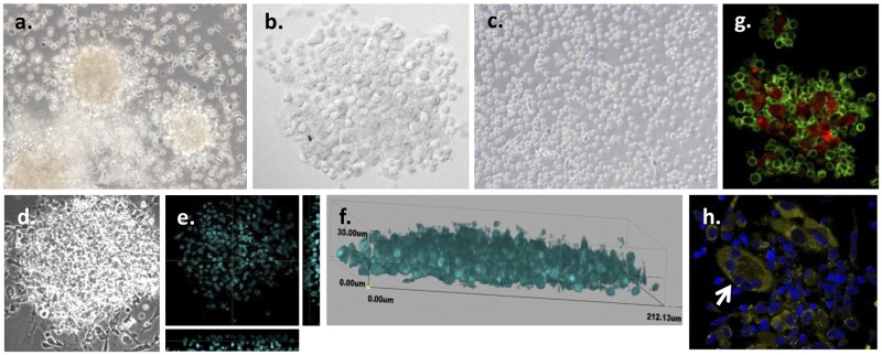
FIG 1 .
In vitro granuloma-like structures are formed by M. tuberculosis infection of PBMCs from LTBI individuals. (a) PBMCs obtained from LTBI individuals were infected with M. tuberculosis H37Rv (MOI 1:1), resulting in the formation of granulomas by day 7 postinfection. (b) Higher magnification of the granulomas by confocal microscopy. (c) No formation of granulomas was observed in uninfected PBMCs obtained from LTBI individuals for up to 12 days postinfection. (d to f) Confocal microscopy images of the granulomas at day 7 postinfection revealed multicellular, multilayered structures containing approximately 4 to 8 cell layers. (d) Differential inference contrast (DIC) image. (e) Image of transverse and straight sections by orthogonal view. (f) Three-dimensional (3D) view image. Nuclei were stained with DAPI (cyan). (g) Granuloma-like structures include macrophages (CD11b+, red) and T cells (CD3+, green). (h) Confocal microscopy images of the granulomas at day 7 postinfection revealed the presence of multinucleated giant cells (CD11b++, yellow; white arrow). Nuclei were stained with DAPI (dark blue). Representative images from n = 12 experiments are shown. The images in panels a and c are shown with ×40 magnification; the remaining images are ×60.

