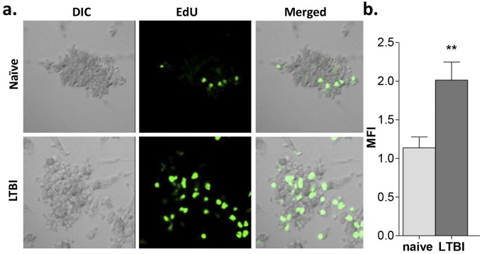
FIG 4 .
The proliferative activity of granuloma-like structures from LTBI individuals is significantly greater than that seen with naive individuals. PBMCs obtained from LTBI and naive individuals were infected with M. tuberculosis H37Rv (MOI 1:1) for up to 7 days, and cell proliferation was determined by EdU assay. (a) Confocal microscopy images of the granulomas at day 7 postinfection revealed greater proliferation (green) in granulomas from LTBI individuals than in those from naive individuals. (b) Quantification of the cellular fluorescence shown by calculation of MFI values. Naive individuals, light grey bars; LTBI individuals, dark grey bars. Uninfected cells were used as controls. All images are ×40. n = 3. **, P < 0.01 (by t test).

