. 2015 Feb 3;4:e04000. doi: 10.7554/eLife.04000

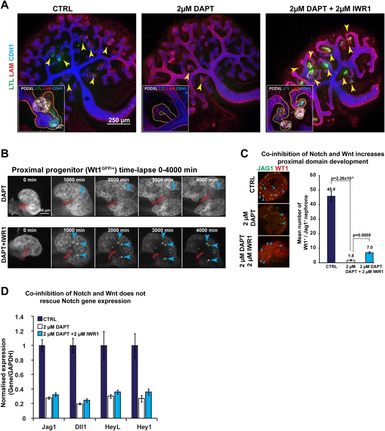
Figure 7. Altered β-catenin activity rescues the loss of Notch.

(A) Kidneys treated with DAPT and DAPT/IWR1 and stained for LTL, β-laminin, Cdh1, and Podxl–arrowheads indicate LTL-positive nephrons, inserts show magnified nephrons with Podxl staining for podocytes, yellow line outlines nephron tubules. (B) Time-lapse analysis of Wt1+/GFP kidneys treated with DAPT and DAPT + IWR1–arrowheads show GFPHIGH structures in developing proximal segments, red dashed line indicates ureteric bud positions (UB). (C) Structures positive for Jag1 and Wt1 in treated kidneys–arrowheads indicating double-positive structures. (D) qRT-PCR data for Notch target genes (Jag1, Dll1, Heyl, Hey1). All error bars indicate SEM.

DOI: http://dx.doi.org/10.7554/eLife.04000.031

Figure 7.

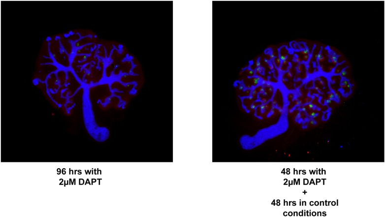
Figure 7—figure supplement 1. Rescue/reversals experiments for kidneys treated with 2 µM DAPT.

Figure 7—figure supplement 1.

Kidneys were cultured for 96 hr in DAPT or for 48 hr in DAPT followed by another 48 hr in control medium. Kidneys were stained for Wt1, Jag1, and Cdh1 to display nephron formation and overall morphology. Antibody stains indicated.
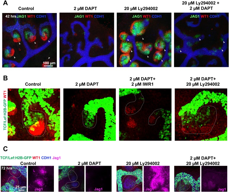
Figure 7—figure supplement 2. Altered β-catenin activity or PI3K signalling rescues the loss of Notch.

Figure 7—figure supplement 2.

Data from kidneys treated with 2 µM DAPT and in combination with 20 µM Ly294002 or 2 µM IWR1. (AB) TCF/Lef:H2B-GFP nephrons stained for WT1, JAG1, and CDH1. WT1 and JAG1 stains are not optimally compatible with the PFA fixation required to preserve the GFP signal. Thus, wild-type kidneys were also treated and stained for WT1 JAG1 and CDH1—shown in (C). Jag1+ structures indicated with arrowheads. Stains, scales, and treatments as specified.