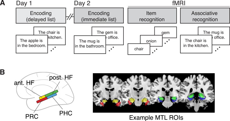
Figure 1. Task design and regions of interest.
(A) Overview of the experimental design. Here, all fMRI analyses are conducted on data from the item recognition phase. (B) ROIs included in the main analyses, including the anterior hippocampus (ant. HF), posterior hippocampus (post. HF), perirhinal cortex (PRC), and parahippocampal cortex (PHC). Coronal MRI slices show manually-traced ROIs from a representative subject, resliced to functional resolution and warped into MNI space for display on a template brain.

