. 2014 Dec 31;3:e03751. doi: 10.7554/eLife.03751

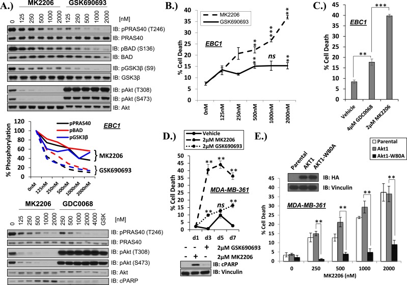
Figure 1. Differential sensitivity of human cancer cells to different classes of AKT inhibitors.

(A) Effect of AKT inhibitors on phosphorylation of AKT and AKT protein substrates. EBC1 cells were treated with the indicated doses of MK2206 and GSK690693 (top), or GDC0068 (bottom) for 24 hr. Treated cells were lysed and analyzed by immunoblot with the indicated antibodies. Phosphorylation of PRAS40, BAD, and GSK3β was quantified by image densitometry (middle). (B) Allosteric AKT inhibitor MK2206 induces more cell death than ATP-competitive inhibitor GSK690693 in EBC1 human lung cancer cells. Cells were treated with drug or vehicle for 96 hr. Cell death was determined by the trypan blue method. Error bars denote standard error of the mean (t-test *p ≤ 0.05, **p ≤ 0.01, treatment vs vehicle). (C) Allosteric AKT inhibitor MK2206 induces more cell death than ATP-competitive inhibitor GDC0068. Experimental conditions were as in Figure 1B. (D) Allosteric AKT inhibitor MK2206 induces more cell death than the ATP-competitive inhibitor GSK690693 in MDA-MB-361 human breast cancer cells. Cell death was assessed on day 1, day 3, day 5, and day 7 following treatment with vehicle or 2 µM of MK2206 or 2 µM of GSK690693(top) (t-test *p ≤ 0.05,**p ≤ 0.01, treatment vs vehicle). An additional plate from each treatment group was lysed 24 hr after drug treatment and analyzed by Western blot with the indicated antibodies (bottom). (E) Suppression of MK2206-induced cell death by a drug-resistant allele of AKT1. MDA-MB-361 cells were stably transduced with HA-tagged wild-type AKT1 or AKT1-W80A and treated with 2 µM MK2206 for 96 hr. Cell death was assessed as above. Expression of the transgenes was confirmed by immunoblot using an HA antibody, and loading was controlled with a vinculin antibody (inset).

DOI: http://dx.doi.org/10.7554/eLife.03751.003

Figure 1—source data 1. Contains source data for Figure 1 and all accompanying Figure 1—figure supplements.
elife03751s001.xlsx (42.7KB, xlsx)
DOI: 10.7554/eLife.03751.004

Figure 1.

Figure 1—figure supplement 1. MK2206 and GSK690693 cause sustained suppression of AKT-dependent BAD phosphorylation.
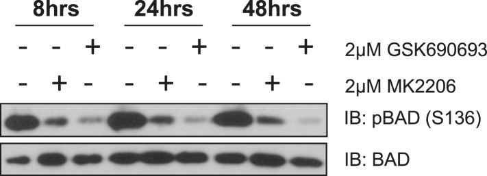
Figure 1—figure supplement 1.

EBC1 cells were treated with either 2 µM MK2206 or 2 µM GSK690693 and lysates were collected at the indicated times. Ser136 BAD phosphorylation was analyzed by immunoblot as indicated.

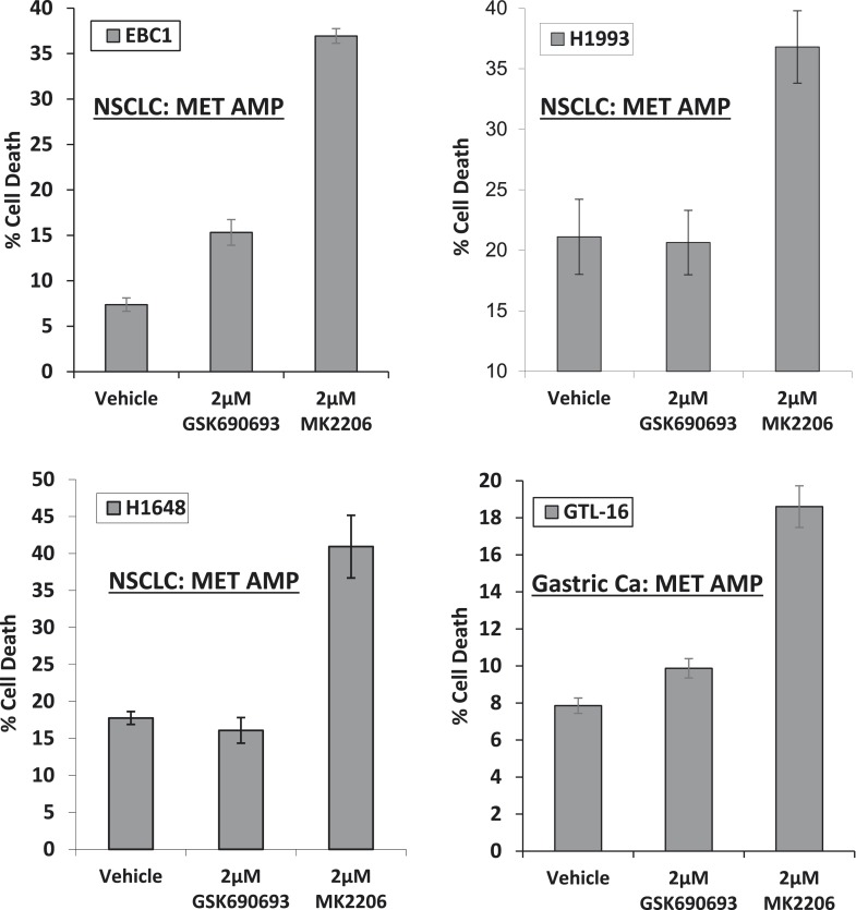
Figure 1—figure supplement 2. MET-amplified cancer cell lines die in response to an allosteric but not an ATP-competitive AKT inhibitor.

Figure 1—figure supplement 2.

EBC1, H1993, H1648, and GTL6 cells were treated with vehicle, 2 µM GSK690693, or 2 µM MK2206 for 96 hr. Cell death was assessed by the trypan blue method as detailed in the ‘Methods’ section.

Figure 1—figure supplement 3. Breast cancer cell lines with HER2 amplification and/or activating PIK3CA-mutations die in response to an allosteric but not an ATP competitive AKT inhibitor.

Figure 1—figure supplement 3.

MDA-MB-361, MCF7, and BT-474 breast cancer cells were treated with the indicated doses of GSK690693 or MK2206 for 96 hr. Cell death was assessed by the trypan blue method.

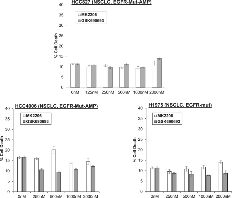
Figure 1—figure supplement 4. Non-small-cell lung cancer (NSCLC) cell lines with EGFR gene amplification and/or activating EGFR mutations are resistant to cell death induction by AKT inhibitors.

Figure 1—figure supplement 4.

HCC827 (EGFR-Δ74-750/AMP) (top right), HCC4006 (EGFR-Δ747-749/AMP) (bottom left), and H1975 (EGFR-T790M-L858R) (bottom right) lung cancer cells were treated with the indicated doses of GSK690693 or MK2206 for 96 hr. Cell death was assessed by the trypan blue method.

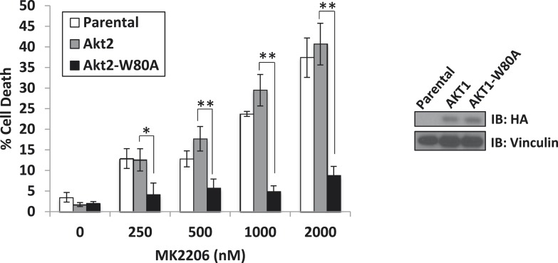
Figure 1—figure supplement 5. A drug-resistant allele of AKT2 suppresses MK2206-induced cell death.

Figure 1—figure supplement 5.

MDA-MB-361 cells were stably transduced with HA-tagged wild-type AKT2 or AKT2-W80A and treated with the indicated concentrations of MK2206 for 96 hr. Cell death was assessed by the trypan blue method (left). Expression of the transgenes was confirmed by immunoblot using an anti-HA antibody, and anti-vinculin was used to control for loading (right).

Figure 1—figure supplement 6. W80A mutation renders AKT1 and AKT2 resistant to MK2206.

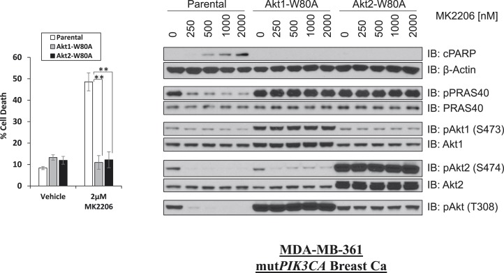
Figure 1—figure supplement 6.

MDA-MB-361 cells were stably transduced with HA-tagged AKT1-W80A or AKT2-W80A and treated with 2 µM MK2206 for 96 hr. Cell death was assessed by the trypan blue method (left). Parental MDA-MB-361 cells or MDA-MB-361 cells stably expressing AKT1-W80A or AKT2-W80A were treated with the indicated doses of MK2206 for 24 hr and lysed. To assess drug effects, lysates were subjected to immunoblot analysis with the indicated antibodies (right).

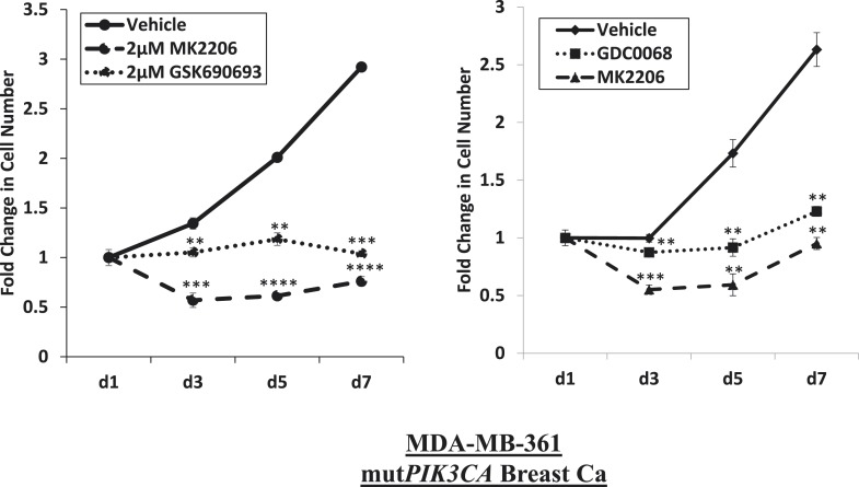
Figure 1—figure supplement 7. Near-complete inhibition of tumor cell proliferation by both allosteric and ATP-competitive inhibitors of AKT.

Figure 1—figure supplement 7.

PIK3CA-mutant MDA-MB-361 cells were treated with vehicle, 2 µM MK2206, or 2 µM GSK690693 (left panel), or vehicle, 2 µM MK2206, or 4 µM GDC0068 (right panel). The number of viable cells was counted on day 1, day 3, day 5, and day 7.

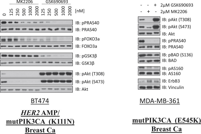
Figure 1—figure supplement 8. Comparison of AKT kinase inhibition by GSK690693 and MK2206.

Figure 1—figure supplement 8.

BT474 cells (left) and MDA-MB-361 cells (right) were treated with the indicated doses of either MK2206 or GSK690693 for 24 hr and lysed. Lysates were analyzed by immunoblot with the indicated antibodies.

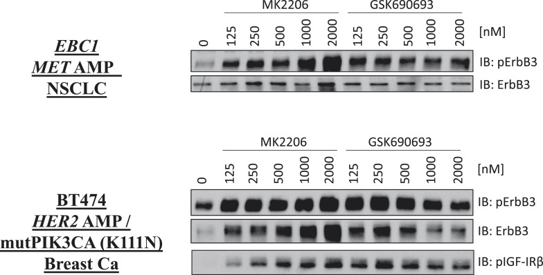
Figure 1—figure supplement 9. De-inhibition of ErbB3 and p-IGF-IRβ by GSK690693 and MK2206.

Figure 1—figure supplement 9.

EBC1 and BT474 cells were treated with the indicated doses of MK2206 and GSK690693 for 24 hr and lysates were analyzed by Western blot with the indicated antibodies.