Figure 2.
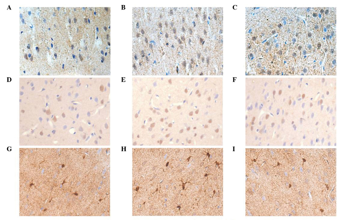
(A–C) Immunohistochemical staining for integrin αv in the mPFC. (A) Control, (B) SPS 4 day, and (C) SPS 7 day groups. (D–F) Immunohistochemical staining for vinculin in mPFC. (D) Control, (E) SPS 4 day, and (F) SPS 7 day groups. (G–I) Immunohistochemical staining for connexin43 in the mPFC. (G) Control, (H) SPS 4 day, and (I) SPS 7 day groups. mPFC, medial prefrontal cortex; SPS, single-prolonged stress.

