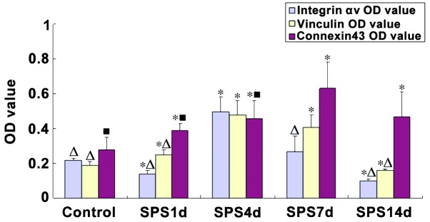
Figure 3.

Quantitative analysis of the average ODs of integrin αv, vinculin and connexin43 as determined by immunohistochemistry in the medial prefrontal cortex. Data are represented as the mean ± standard deviation. *P<0.05 compared with control, ΔP<0.05 compared with SPS 4 day, =P<0.05 compared composed with SPS 7 day. SPS, single-prolonged stress; OD, optical density.
