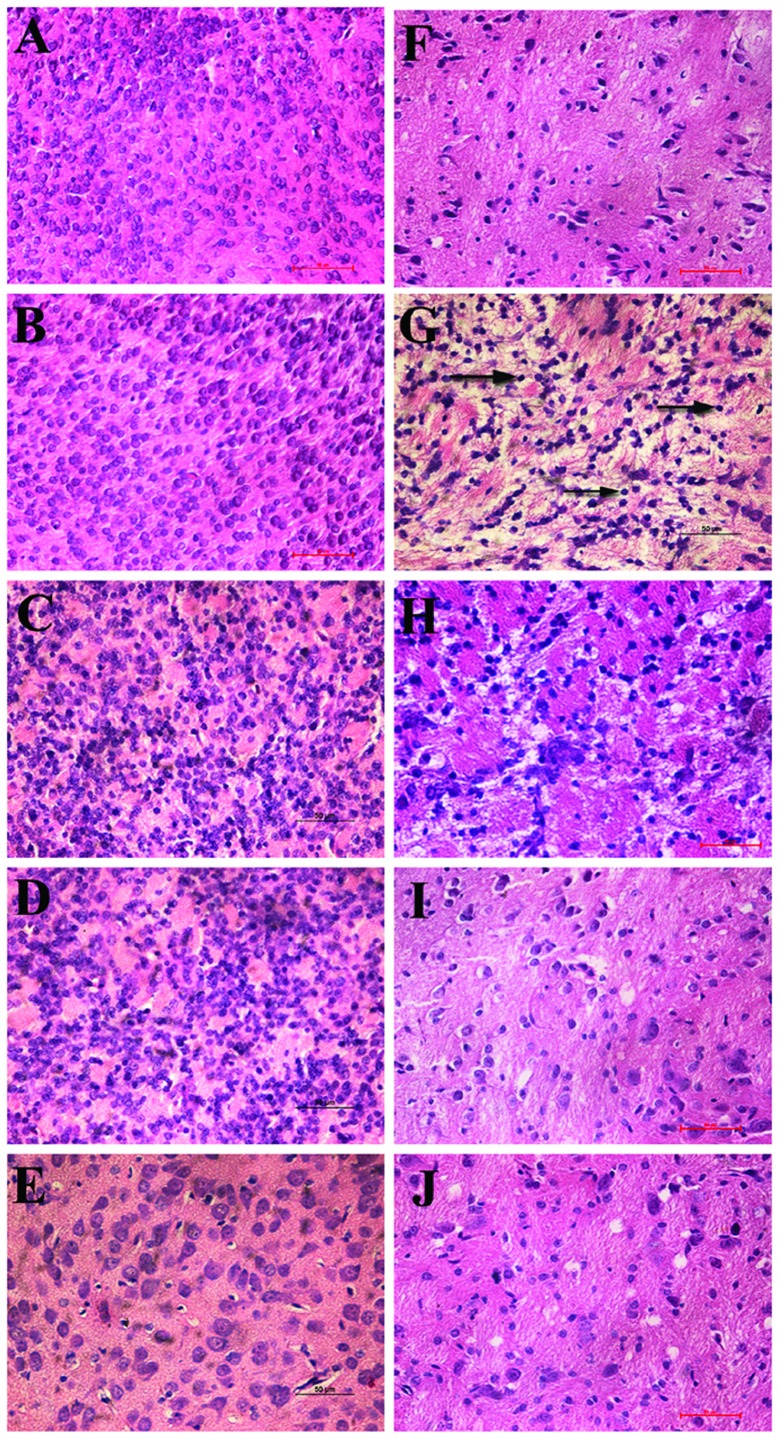
Figure 1.
Malacotic periventricular brain tissues caused by hypoxia-ischemia (HI), as observed with hematoxylin and eosin (H&E) staining (magnification, ×400). Brain tissues of the normoxia group exhibited a normal morphology, and uniform, intensive cell distribution at all time-points (A–E). In the HI group, karyopyknosis was observed in brain tissues, with necrosis of a few cells at day 1 (F). At day 3, the focus of malacia began to appear in the alba, and necrosis was observed in a high number of cells, along with a cribriform change (G). At day 7, the number of cystic necroses was reduced compared to day 3, when the necroses first appeared (H). At days 14 and 21 (I,J), the number of cystic necroses was smaller, and cribriform necroses gradually disappeared or were transformed into areas of cluster or punctiform necroses, although brain tissues still had a porous, malacotic structure.

