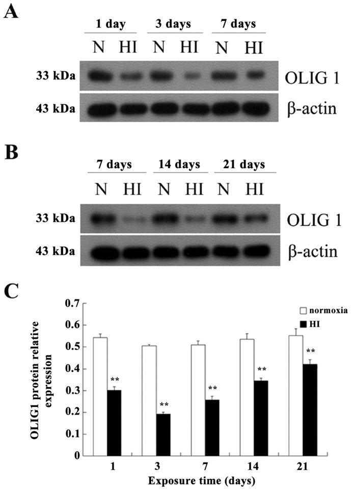
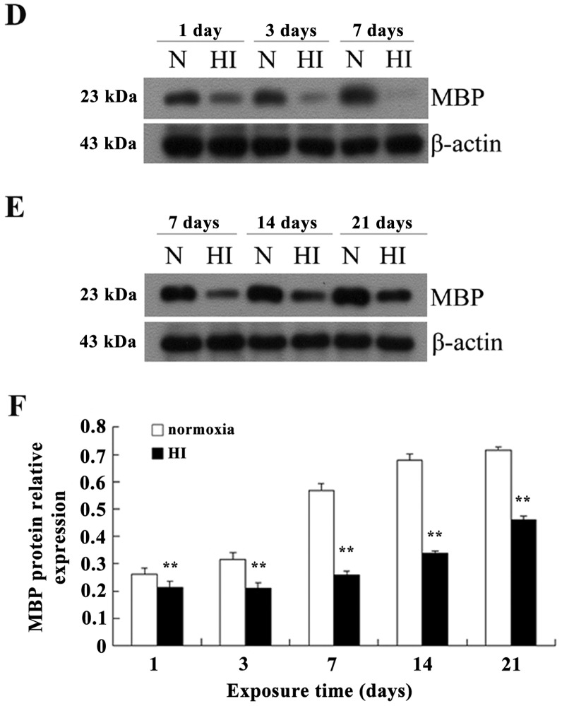
Figure 6.
Decreased expression of OLIG1 and myelin basic protein (MBP) protein in brain tissues following hypoxia-ischemia (HI). In the normoxia (N)group, the expression level of OLIG1 in brain tissues is not significantly different (P>0.05) at different time-points (C). In the HI group, the lowest expression level of OLIG1 is observed at day 3 (A). The expression level of the MBP protein in the normoxia group shows a progressively increasing (P<0.01) trend (C). At 7 days of HI exposure, the MBP expression dramatically declines, and is virtually undetectable (P<0.01) by western blotting (D and E). Increased expression of the OLIG1 and MBP proteins is observed in newborn rats at days 14 and 21. In the HI group, despite this increase, the levels of the OLIG1 and MBP proteins (P<0.01) do not reach the expression levels observed in the normoxia group (B–C and E–F). The relative expression level of OLIG1 and MBP proteins (P<0.01) in the brain tissue was calculated by the mean value of the relative optical densities (C and F). Means ± standard deviation (SD) values are shown. **P<0.01 vs. the normoxia group. All experiments were performed independently 4 times.


