Figure 1.
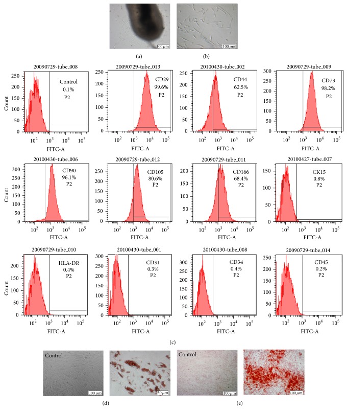
Isolation and characterization of hHFMSCs. (a) The hHFMSCs, resembling typical fibroblast-like cells, migrated out from the hair follicles (original magnification ×100). (b) hHFMSCs from passage 3 (original magnification ×100). (c) Flow cytometric analysis of cell surface markers on hHFMSCs. 2 × 10⁵ cells were incubated with primary antibodies, followed by incubation with a secondary FITC-labeled antibody. Controls were incubated with secondary antibody only. Percentages indicate the fraction of cells that stained positive. (d) Adipogenic differentiation of hHFMSCs. Compared to noninduced control (original magnification ×100), induction after 3 weeks, the number of intracellular lipid droplets was developed and increased and was detected by Oil-red O staining (original magnification ×200). (e) Osteogenic differentiation of hHFMSCs. Calcium nodules were formed after induction for 4 weeks and were demonstrated by Alizarin red S staining (original magnification ×100).

