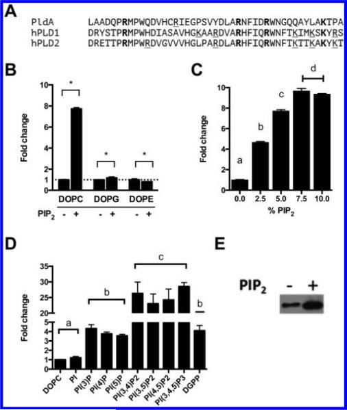
Figure 4.
Phosphoinositides stimulate PldA activity and promote lipid binding in vitro. (A) Alignment of putative PI(4,5)P2 binding domain of PldA and PI(4,5)P2 binding domain of human PLD1 and PLD2. (B) Brain PI(4,5)P2 stimulates PC substrate hydrolysis by PldA. Statistical analysis performed using Student's t test. *p < 0.01. (C) Brain PI(4,5)P2 stimulation of DOPC hydrolysis is concentration dependent. Statistical analysis performed using one-way ANOVA with Tukey's post hoc test. Groups differing (p < 0.01) have different letters. (D) Phosphoinositides stimulate DOPC hydrolysis. All phosphoinositides were 36:2, except for brain PI(4)P. Statistical analysis performed using one-way ANOVA with Tukey's post hoc test. Groups differing (p < 0.01) have different letters. (E) Brain PI(4,5)P2 increases DOPC liposome binding of PldA. All experiments performed in triplicate. Shown are representative data. All mean values are shown ± SEM (n = 3).

