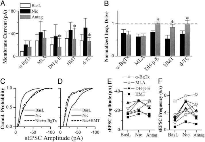
FIG. 3.
Cellular level effects of Nic and Nic Antag in preBötC inspiratory neurons voltage-clamped at −60 mV. The effects of α-BgTx (n = 4) or MLA (n = 4) on Nic-induced tonic inward current (A) and decrease in amplitude of phasic inspiratory drive current (B) were not significant; DH-β-E (n = 5), HMT (n = 4), and d-TC (n = 4) reversed these nicotine-induced responses. The amplitude of phasic inspiratory drive current was normalized by the values prior to application of nicotine (baseline: BasL). One-way repeated-measures ANOVA for each group and posthoc analysis based on Tukey were used for A and B. *, statistically significant. C: α-BgTx did not reduce nicotine-induced increase in amplitude of spontaneous EPSCs (sEPSCs). Cumulative (Cumul) histogram from 1 representative neuron. D: HMT reversed the Nic-induced increase in amplitude of sEPSCs. Summaries of the effects of α-BgTx, MLA, DH-β-E, and HMT on the Nic-induced changes in amplitude (E) and frequency (F) of sEPSCs. sEPSCs were recorded for 1–2 min, and 100–600 events were collected in each condition for each neurons. Each symbol indicates 1 neuron. Refer to results for statistical analyses for C–F.

